Plain Radiograph/X-ray
What is a plain radiograph/X-ray? This is the most simple, cost-effective and readily accessible imaging technique available. It provides excellent imaging details…
Read more
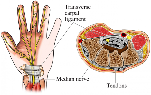
The carpal tunnel is located in the wrist, and is formed by the bones of the wrist and the transverse carpal (wrist) ligament. Through this tunnel pass the median nerve, flexor tendons (which help you to curl your fingers) and tendon sheaths (the coverings of the tendons).
Carpal tunnel syndrome (also known as median nerve compression) is a common cause of symptoms in the hand and wrist.
Ultrasound is used to take pictures or images of the wrist to confirm or exclude if carpal tunnel syndrome is present. Ultrasound is good at assessing if there is swelling of the median nerve and also assessing the adjacent structures (such as joints and tendons), which might be irritating or compressing the nerve.
If carpal tunnel syndrome is found to be present, ultrasound is used to guide the placement of a needle into the carpal tunnel to inject a small dose of corticosteroid (or ‘steroid’) and local anaesthetic medication.

The carpal tunnel lies between the wrist bones and the flexor retinaculum (ligament), and is filled by tendons and the median nerve. An injection is targeted to fill the space immediately adjacent to the median nerve at the ‘roof’ of the tunnel.
You would be referred if you have symptoms that your doctor believes might be carpal tunnel syndrome. Your doctor might have tried simple measures, such as rest, changing activities and splinting (putting something around the wrist and hand to keep them from moving). If these have not been successful in relieving your symptoms, an ultrasound of the carpal tunnel might be requested with a view to possible injection.
Carpal tunnel syndrome is caused by swelling or compression of the median nerve as it passes through the carpal tunnel from the forearm into the hand. The carpal tunnel is a narrow space, and swelling within or around it can compress the median nerve and lead to the development of symptoms, such as tingling and numbness and pain, on the thumb side of the hand up to the ring finger. Muscle weakness can also occur. There are numerous causes of carpal tunnel syndrome, some are due to associated medical problems (see websites at the end of this section). Tendinopathy (swelling of the tendons), synovitis (swelling and inflammation of the tendon sheaths) or joint swellings around the carpal tunnel can also cause symptoms of carpal tunnel syndrome.
The corticosteroid injection should reduce inflammation or swelling in and around the median nerve, the flexor tendons and the tendon sheaths, which in turn should relieve your symptoms.
No specific preparation is needed. You should take any previous X-rays or scans to the appointment. You might feel numbness or pins and needles in your hand for up to 1 hour after the injection, and driving is not recommended during this period. It is generally advised that you bring someone with you who can drive you home.
When you make your appointment for the ultrasound and injection, you need to let the radiology clinic or department know if you are taking any blood thinning medication, particularly warfarin.
Blood thinning medications might need to be stopped for a period of days, or your normal dose reduced, before this procedure is carried out. The radiology clinic or department or your own doctor will give you specific instructions about whether you need to stop or reduce the medication and when to restart the medication. These drugs are usually taken to prevent stroke or heart attack, so it is very important that you do not stop taking them without being instructed to do so by your doctor or the radiology practice, or both.
A blood test might be required to check your blood clotting before the procedure.
Continue with pain medication and other medications as usual.
You will be taken into the scanning room by the sonographer (ultrasound technician). You will either be lying on a scanning bed or sitting down with your hand on a table or bed in a comfortable position. The sonographer will apply gel over your wrist and take images using ultrasound. These images will then be shown to the radiologist (specialist doctor) who will discuss them with you and might take some further images.
If carpal tunnel syndrome is confirmed, and the radiologist recommends an injection, the procedure will be explained to you. You will be able to ask any questions at this time. The skin over your wrist is cleaned with antiseptic liquid. A small needle is passed through your skin directly into the carpal tunnel using ultrasound images to guide the placement of the needle. A small amount of corticosteroid (or ‘steroid’) and local anaesthetic (usually just a few millilitres) is then injected, and the needle removed. Most people are surprised by how quick and easy the procedure is.
The radiologist will give you advice for after the injection. The wrist and hand should generally be rested completely for 6 hours, followed by minimal use for between 1 and 3 days.
Immediately after the injection, you might have numbness in your hand from the local anaesthetic. It is recommended that you do not drive until the numbness has settled or have someone drive you home after the procedure.
The most common after effect is a temporary increase of your symptoms over 1, 2, or even 3 days. The corticosteroid does not start working for at least 24 hours and sometimes up to 7 days.
Symptoms from nerves generally take longer to respond to corticosteroid than symptoms relating to muscles or joints. During this time, the normal symptoms might continue or, occasionally, are worse. A major flare of symptoms generally indicates a local reaction to the injected medication or to having the needle. Anti-inflammatory medication, rest (use of a splint) and the application of cold packs is recommended. If the reaction is persistent, then you should seek medical attention, as it might be an infection, although this is unlikely.
Sometimes you might have general symptoms related to absorption of the corticosteroid into the circulation. This usually only occurs when larger doses are used or in some people who are more sensitive to corticosteroids. In diabetics, this absorption might increase the blood sugar levels (BSL) for a few days, and BSL should generally be checked several hours after the procedure.
Slight bruising and bleeding might occur around the wrist as a result of the needle.
If your doctor has requested a full ultrasound scan, this can take up to 40 minutes. The injection procedure itself rarely takes more than 5 minutes, but with the preparation (scanning, marking, cleaning the skin, etc.) it can take up to 15 minutes.
This is a very safe procedure with few significant risks, but occasionally problems are experienced.
Sometimes the ultrasound is carried out for diagnostic reasons, if your doctor is uncertain as to the cause of your symptoms.
The aim of the injection is to provide symptom relief. A good response to the injection confirms the diagnosis of carpal tunnel syndrome. A lack of response (no improvement in symptoms) indicates the pain is unlikely to be due to carpal tunnel syndrome. Although having the injection might not result in any improvement in your symptoms, it can be helpful information for your doctor, as it means that other possible causes need to be investigated.
The relief of symptoms might last a few weeks or several months. Occasionally, (usually when the condition is recent or due to recent overuse), the symptoms resolve completely.
A sonographer (or sometimes a specialist doctor) will carry out the ultrasound and take the images. The injection is administered by a radiologist (specialist doctor). The radiologist will provide a written report, which is sent to your doctor detailing the findings and the procedure.
A carpal tunnel ultrasound and injection is usually carried out in a hospital radiology department or a private radiology or nuclear medicine practice.
The time that it takes your doctor to receive a written report on the test or procedure you have had will vary, depending on:
Please feel free to ask the private practice, clinic or hospital where you are having your test or procedure when your doctor is likely to have the written report.
It is important that you discuss the results with the doctor who referred you, either in person or on the telephone, so that they can explain what the results mean for you.
Nerve conduction tests (involving electrically activating nerves and measuring the responses) and magnetic resonance imaging (MRI) can also be used for diagnosis. Anti-inflammatories, simple analgesia, wrist splints and rest can all provide relief in some patients.
Depending on the possible underlying cause and contributing factors of the carpal tunnel syndrome, other medical management might be advised.
Surgical decompression (open or key-hole) is commonly carried out for carpal tunnel syndrome.
US National Library of Medicine
www.ncbi.nlm.nih.gov/pubmedhealth/PMH0001469/
Better Health Channel
www.betterhealth.vic.gov.au/bhcv2/bhcarticles.nsf/pages/Carpal_tunnel_syndrome
Page last modified on 26/7/2017.
RANZCR® is not aware that any person intends to act or rely upon the opinions, advices or information contained in this publication or of the manner in which it might be possible to do so. It issues no invitation to any person to act or rely upon such opinions, advices or information or any of them and it accepts no responsibility for any of them.
RANZCR® intends by this statement to exclude liability for any such opinions, advices or information. The content of this publication is not intended as a substitute for medical advice. It is designed to support, not replace, the relationship that exists between a patient and his/her doctor. Some of the tests and procedures included in this publication may not be available at all radiology providers.
RANZCR® recommends that any specific questions regarding any procedure be discussed with a person's family doctor or medical specialist. Whilst every effort is made to ensure the accuracy of the information contained in this publication, RANZCR®, its Board, officers and employees assume no responsibility for its content, use, or interpretation. Each person should rely on their own inquires before making decisions that touch their own interests.